Most patients — adults and parents alike — have quietly wondered whether dental X-rays are truly necessary or just a routine revenue line for dental practices. It’s a fair question, and it deserves a straight answer.
The short answer: dental X-rays are one of the most diagnostically valuable tools in dentistry — but not every patient needs them at every visit, and costs vary significantly depending on type, location, and insurance coverage.
This guide will walk you through exactly why dentists recommend X-rays, what how much do dental X-rays cost really means across different types, what insurance typically covers, and how to make an informed decision for yourself or your child.
According to the American Dental Association, dental X-rays are most effective when used selectively based on individual risk — not as a one-size-fits-all routine.
What Are Dental X-Rays?
Dental X-rays (also called dental radiographs) are diagnostic images that allow dentists to see structures that are invisible to the naked eye — including the roots of teeth, the jawbone, developing permanent teeth in children, and decay forming between teeth or beneath existing fillings.
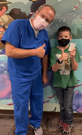
Our providers are specifically trained in interpreting pediatric dental radiographs — a skill set that requires specialty education beyond general dentistry. Dr. Brad Dawson, who holds dual post-doctorate degrees in both Pediatric Dentistry and Orthodontics from Indiana University, emphasizes that X-rays are especially critical during the mixed dentition years (ages 6–12), when adult teeth are actively developing beneath the gumline, and problems caught early can prevent costly, complex treatment later.
They use a small, controlled amount of ionizing radiation to create images of the internal structures of the mouth. Modern digital X-ray systems have dramatically reduced radiation exposure compared to traditional film X-rays — typically by 70–80%.
The four most common types:
| Type | What It Shows | Common Use |
| Bitewing X-rays | Crowns of upper and lower back teeth | Detecting cavities between teeth |
| Periapical X-rays | Full tooth from crown to root tip | Root infections, abscesses, bone loss |
| Panoramic X-rays | Entire mouth in one image | Jaw development, wisdom teeth, orthodontics |
| Full mouth series | Complete set of 14–21 images | Comprehensive new patient assessment |
Defining insight: A dental X-ray doesn’t just find cavities — it reveals bone loss, cysts, tumors, impacted teeth, and developmental abnormalities that have no visible symptoms until they become serious and expensive problems.
The U.S. Food and Drug Administration also provides guidance on when dental X-rays are necessary, emphasizing their role in diagnosis when clinical examination alone is not sufficient.
Why Dental X-Rays Are Necessary: What They Catch That Exams Cannot
The most common objection to dental X-rays is simple: “My teeth look fine.” The problem with that reasoning is that most significant dental disease develops where no one can see it.
Here’s what a visual exam alone will miss:
Interproximal decay — cavities that form between teeth are completely invisible without X-rays until they’ve grown large enough to cause visible damage or pain. By then, a simple filling has often become a root canal.
Bone loss from periodontal disease — gum disease that has progressed to affect the supporting bone shows no symptoms until teeth begin to loosen. X-rays reveal bone levels long before clinical symptoms appear.
Root infections and abscesses — an infected tooth root can exist for months without obvious pain, especially in children. A periapical X-ray shows the infection before it spreads to surrounding bone or becomes a systemic health risk.
Developmental abnormalities in children — panoramic X-rays in children reveal whether permanent teeth are forming correctly, whether they are impacted, and whether early orthodontic intervention is needed. None of this is visible by looking in a child’s mouth.
The cost of dental X-rays without insurance is almost always far less than the cost of treating the conditions they catch early.
The dentists at Great Falls Pediatric Dentistry & Orthodontics use advanced digital dental X-rays as a core diagnostic tool — not a formality. According to our clinical team, X-rays allow us to detect decay between teeth, monitor developing permanent teeth, identify bone abnormalities, and plan treatments that simply cannot be accurately developed from a visual exam alone. Our approach is always to use the minimum radiation exposure necessary while still achieving diagnostic precision.
Key Factors That Determine How Often You Need Dental X-Rays
1. Your Individual Cavity and Disease Risk
The American Dental Association provides updated, evidence-based recommendations on dental X-rays and does not support a fixed schedule for all patients.
Higher risk patients — those with a history of frequent cavities, active gum disease, dry mouth, diabetes, or undergoing orthodontic treatment — may need bitewing X-ray cost assessed every 6 months.
Lower risk patients — adults with no recent cavities, healthy gums, and stable dental history — may only need bitewing X-rays every 18–24 months.
A dentist who recommends annual X-rays for every patient regardless of risk profile is not following ADA evidence-based guidelines. A dentist who never recommends them is being negligent.
2. Your Age and Developmental Stage
Children and teenagers need X-rays more frequently than low-risk adults because their mouths are actively changing. Panoramic dental X-ray imaging is particularly valuable for children aged 6–12 to monitor the eruption of permanent teeth and detect early orthodontic issues.
For adults over 50, full mouth dental X-ray series every 3–5 years provide a comprehensive baseline for monitoring bone levels and detecting early-stage oral cancer or cysts.
3. Whether You’re a New Patient
New patient X-rays — typically a full mouth series or panoramic plus bitewings — are standard practice and clinically justified. A dentist cannot responsibly treat a patient they’ve never imaged. It’s equivalent to a physician treating a patient without reviewing their medical history.
4. Existing Dental Work
Patients with multiple fillings, crowns, root canals, or implants need more frequent imaging to monitor the integrity of existing restorations and the bone and tissue surrounding them.
5. Symptoms or Clinical Findings
Any time a patient presents with pain, swelling, sensitivity, or a visible lesion, targeted X-rays are diagnostically necessary — not optional. This is true regardless of when the last set of X-rays was taken.
Common Mistakes Patients Make About Dental X-Rays
Refusing X-rays based on radiation fear. A full set of dental X-rays exposes a patient to approximately 0.005 millisieverts of radiation — equivalent to less than one day of background radiation from the natural environment. The risk is negligible. The diagnostic benefit is substantial.
Assuming “no pain” means no X-rays needed. The most dangerous dental conditions — bone loss, root infections, early-stage oral cancer — are frequently painless in their early stages. Pain is a late-stage symptom, not a reliable indicator of disease.
Skipping X-rays to save money at a new dental office. Bringing X-rays from a previous dentist is a legitimate cost-saving measure. X-rays taken within the past 12–24 months may be transferable. Ask your previous office for digital copies before your new patient appointment.
Believing that are dental X-rays covered by insurance is always a yes. Coverage varies significantly by plan. Some plans cover bitewing X-rays annually; others cover them every 12–18 months. Panoramic X-rays may be covered only once every 3–5 years. Always verify before your appointment.
Average Price of Dental X-Rays in 2026: What to Expect
The cost of dental X-rays without insurance varies by type, geographic location, and dental practice. Here is a realistic breakdown based on current market data:
| X-Ray Type | Average Cost (No Insurance) | What’s Included |
| Bitewing X-rays (2–4 images) | $25–$150 | Cavity detection between back teeth |
| Periapical X-rays (per tooth) | $25–$50 | Root and bone around individual tooth |
| Panoramic X-ray | $100–$250 | Full jaw, all teeth in one image |
| Full mouth series | $150–$400 | Complete 14–21 image comprehensive set |
| Cone beam CT (CBCT) | $250–$600 | 3D imaging for implants/complex cases |
The average price of dental X-rays in 2026 has increased modestly compared to prior years, driven primarily by equipment upgrade costs in practices transitioning to digital and 3D imaging systems.
With insurance: Most dental insurance plans cover bitewing X-rays at 100% as part of preventive care. Panoramic and full-mouth series are typically covered at 80% after deductible, subject to frequency limitations.
Cheap dental X-rays near me searches often surface dental schools, community health centers, and Federally Qualified Health Centers (FQHCs) — all of which provide significantly reduced-cost dental services, including X-rays, for uninsured or underinsured patients.
Are Dental X-Rays Covered by Insurance? A Realistic Guide
The short answer: usually yes, partially, with conditions.
What most plans cover:
- Bitewing X-rays: 100%, once every 12–18 months
- Panoramic X-rays: 50–80%, once every 3–5 years
- Full mouth series: 50–80%, once every 3–5 years
- Periapical X-rays: Often covered when diagnostically necessary
What affects your coverage:
- Plan type (DHMOs tend to have stricter frequency limits than PPOs)
- Whether the dentist is in-network
- Whether you’ve met your annual deductible
- Your plan’s benefit year reset date
Always call your insurance provider before an appointment and ask specifically: “What dental X-rays are covered under my plan, and how frequently?” This 5-minute call can save you $100–$300.
Voice Search Questions Answered
What are dental X-rays?
Dental X-rays are diagnostic images that allow dentists to see tooth decay, root infections, bone loss, and developmental issues that are completely invisible during a visual examination. They use a small, controlled dose of radiation and are a standard tool in both preventive and diagnostic dentistry.
Are dental X-rays safe?
Yes. Modern digital dental X-rays use extremely low radiation doses — comparable to less than one day of natural background radiation. The diagnostic benefit of detecting hidden decay, infections, or bone loss far outweighs the negligible radiation exposure for the vast majority of patients.
When should I get dental X-rays?
Frequency depends on your individual risk profile. Higher-risk patients — those with active decay, gum disease, or complex dental history — may need bitewing X-rays every 6 months. Low-risk adults may only need them every 18–24 months. New patients typically need a baseline series regardless of recent history.
How do dental X-rays work?
A small sensor or film is placed inside the mouth and a low-dose X-ray beam passes through the teeth and jaw. Dense structures like enamel and bone absorb more radiation and appear white on the image; softer tissues and decay appear darker. Digital sensors capture the image instantly without chemical processing.
How much do dental X-rays cost without insurance?
Bitewing X-rays typically cost $25–$150. A panoramic X-ray runs $100–$250. A full mouth series ranges from $150–$400. Costs vary by location and practice type. Community health centers and dental schools offer significantly reduced rates for uninsured patients.
Dental X-Ray Checklist: What Every Patient Should Know
✔ Ask your dentist to explain which X-rays are recommended and why ✔ Provide copies of recent X-rays when switching dental providers ✔ Verify your insurance coverage and frequency limits before your appointment ✔ Ask whether the practice uses digital X-rays (lower radiation, instant results) ✔ Understand that children typically need X-rays more frequently than low-risk adults ✔ Never skip X-rays purely based on radiation fear — the dose is clinically negligible ✔ Ask about panoramic X-rays for children aged 6–12 to monitor development ✔ Inquire about dental school or FQHC options if cost is a barrier ✔ Know that pain-free does not mean X-ray-free — most serious issues have no early symptoms ✔ Keep a personal record of your X-ray history and dates for future reference
Conclusion: The Real Cost of Dental X-Rays Is What You Pay When You Skip Them
How much do dental X-rays cost is the question patients ask. The more important question is: how much does it cost when the decay, infection, or bone loss they would have caught goes undetected for another year?
A $75 set of bitewing X-rays that catches an early cavity saves a $1,200 root canal. A panoramic X-ray that identifies an impacted permanent tooth in a 9-year-old saves years of complex orthodontic treatment. A periapical X-ray that reveals an abscess prevents a potentially life-threatening spreading infection.
Dental X-rays are not a revenue tool. They are a diagnostic necessity — when recommended appropriately, based on your individual risk, by a dentist following evidence-based guidelines.
“The most expensive dental X-ray is the one that wasn’t taken.”
If it’s been more than 18 months since your last set, or your child hasn’t had age-appropriate imaging, that’s the only number that matters right now.
Want to understand exactly which X-rays your child needs and when? Speak with a pediatric dental specialist who follows AAPD imaging guidelines. You can also review the ADA’s evidence-based clinical recommendations for dental radiographs or the American Academy of Pediatric Dentistry’s imaging guidelines for age-specific guidance.
Frequently Asked Questions
Q: How often should children get dental X-rays?
A: The AAPD recommends frequency based on individual cavity risk. High-risk children may need bitewing X-rays every 6 months; lower-risk children every 12–24 months. Panoramic X-rays are typically recommended around age 6–7 and again in early adolescence to monitor development.
Q: Can I request to skip dental X-rays?
A: You can decline X-rays, and a dentist must respect your decision. However, understand that doing so limits diagnostic capability. Some practices will ask you to sign an informed refusal form acknowledging that certain conditions may go undetected without imaging.
Q: Are digital X-rays better than traditional film X-rays?
A: Digital X-rays use 70–80% less radiation than traditional film, produce instant images, allow for digital storage and easy transfer between providers, and can be enhanced on-screen for better diagnostic accuracy. They are now the standard of care in modern dental practices.
Q: What is a panoramic dental X-ray used for?
A: A panoramic X-ray captures the entire mouth — all teeth, both jaws, and surrounding structures — in a single image taken from outside the mouth. It is used to assess wisdom tooth development, detect jaw pathology, plan orthodontic treatment, and evaluate overall dental development in children.
Q: Do dental X-rays detect oral cancer?
A: While standard dental X-rays primarily show hard tissue (bone and teeth), they can reveal bone destruction associated with oral cancer. Periapical X-rays and panoramic imaging can show lesions affecting the jaw. Dentists also perform soft tissue visual screenings that complement X-ray imaging for oral cancer detection.
Q: Is it safe to get dental X-rays during pregnancy?
A: The ADA and American College of Obstetricians and Gynecologists state that dental X-rays are safe during pregnancy when clinically necessary, with a lead apron and thyroid collar used for shielding. Delaying necessary X-rays during pregnancy can allow dental disease to progress, which carries its own risks.
Q: Where can I get cheap dental X-rays near me?
A: Dental schools, Federally Qualified Health Centers (FQHCs), and community health clinics offer significantly reduced-cost dental X-rays for uninsured or low-income patients. Search the HRSA Health Center Finder to locate an FQHC near you.